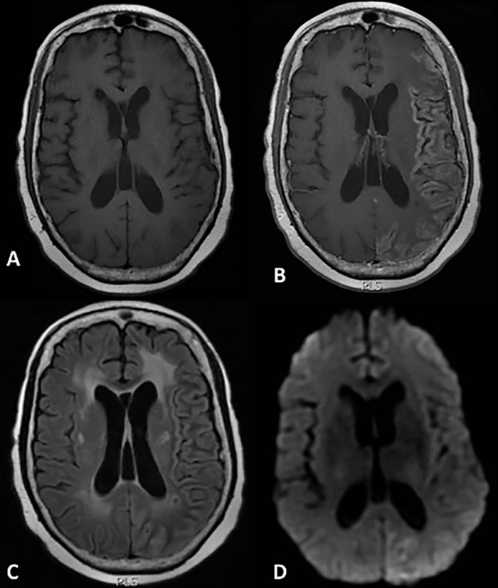
Figure 1.

Magnetic resonance imaging on hospitalization day 9. Axial T1-weighted images through the level of the cerebral hemispheres before (A) and after (B) the administration of gadolinium contrast demonstrate diffuse gyral enhancement throughout much of the left cerebral hemisphere with some sparing of the frontal lobe, with corresponding mildly hyperintense signal on a T2/fluid attenuated inversion recovery image (C) and diffusion restriction on diffusion weighted imaging (D).
