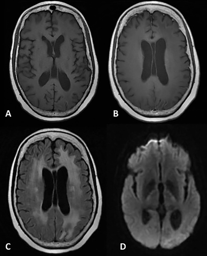
Figure 3.

Magnetic resonance imaging at 5-month follow-up. Axial T1-weighted images before (A) and after (B) the administration of gadolinium contrast demonstrate minimal cortical intrinsic T1 shortening along the left posterior parietal and occipital cortex consistent with laminar necrosis but complete resolution of the postcontrast cortical enhancement. T2/fluid attenuated inversion recovery image demonstrates resolution of the posterior cerebral cortical signal abnormality but new left occipital subcortical hyperintensity, indicative of gliosis and encephalomalacia (C). Diffusion weighted imaging shows resolution of diffusion restriction (D).
