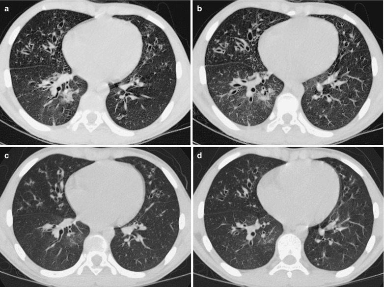
Fig. 22.13.

Influenza A (H1N1) complicated by pneumonia and bronchial dilation. (a, b) CT scanning demonstrates small flake of parenchymal shadow and ground-glass opacity in the anterior lower lobe of the right lung and in the inner basal segment of the right lung, widened bronchial canals, and thickened bronchial wall of both lungs, with signet ring sign. (c, d) By reexamination after treatment for 7 days, CT scanning demonstrates the lesions improved and absorbed
