
Fig. 22.25.
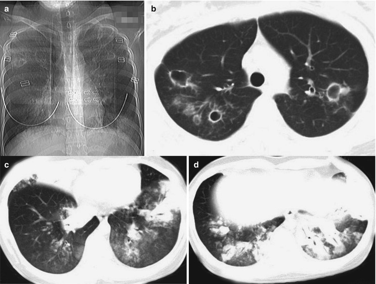
Influenza A (H1N1) complicated by pneumonia and bronchiectasis. (a) X-ray demonstrates flakes of shadows with blurry boundaries in the inner zone of the right middle and lower lung field and the left lower lung field and multiple cavities in both lungs with unclearly defined boundaries. (b) CT scanning demonstrates multiple irregular and thin-walled empty cavities in both upper lobes and surrounding scattering patches of shadows. There are also thickened bronchial wall in the left upper lobe, with widened lumen. (c, d) Multiple consolidations and ground-glass opacities are demonstrated in the right middle lobe, right lower lobe, and inferior lingual segment of the left upper lobe and left lower lobe