
Fig. 22.29.
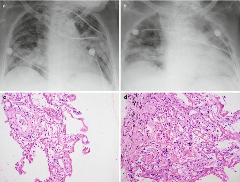
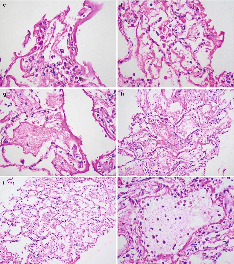
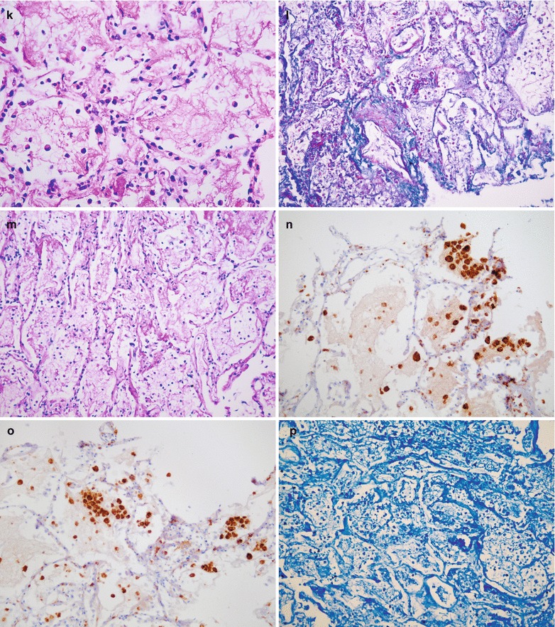
Influenza A (H1N1) complicated by pneumonia. (a) X-ray demonstrates flakes of shadows with increased density in both lung fields that are more obvious in both lower lungs and enlarged blurry hilar shadows. (b) X-ray demonstrates progress of the conditions, with extended range of shadows in both lung fields with high density that are more obvious in the right lower lung and the left lung and enlarged blurry hilar shadows. (c) There are widened interalveolar space, congested alveolar walls, infiltration of neutrophils and plasma cells, and exudations of mononuclear cells, alveolar edema fluid, and celluloses (H&E ×20). (d) Formation of intra-alveolar transparent membrane (H&E ×20). (e) Shedding of alveolar epithelium and exudation of celluloses from some alveolar cavities (H&E ×40). (f) A large quantity of intra-alveolar transparent membranes forms (H&E ×40). (g) Vascular congestion of alveolar walls (H&E ×40). (h) Thinner alveolar walls, occluded vascular vessels, and exudation of celluloses in a large quantity from alveoli (H&E ×20). (i) Following shedding and necrosis of type I epithelial cells, slight hyperplasia of type II epithelial cells occurs (H&E ×20). (j) Exudation of loose celluloses within alveolar cavities (H&E ×40). (k) Exudation of dense celluloses within alveolar cavities (H&E ×40). (l) Exudation of celluloses in a large quantity within alveoli, with no growth of bacteria (Masson staining, ×20). (m) Exudation of celluloses in a large quantity within alveolar cavities (PAS staining × 20). (n, o) Accumulation of macrophages into masses (H&E ×20). (p) A large quantity of inflammatory cell infiltration, with no findings of acid-fast bacilli (H&E ×20)