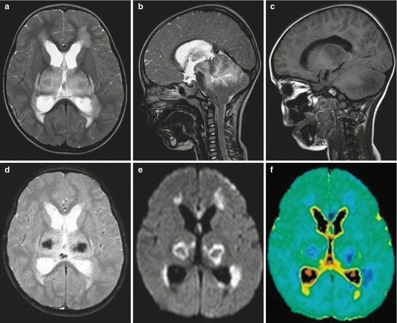
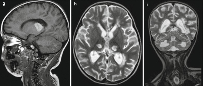
Fig. 22.3.


Influenza A (H1N1) complicated by acute necrotizing encephalopathy. (a) In the acute stage, transverse T2WI demonstrates symmetrically increased signal in the bilateral thalamus and white matter surrounding the anterior and posterior horns of lateral ventricles. (b) Sagittal T2WI demonstrates high signals in the cerebellum and brainstem. (c) Sagittal T1WI demonstrates low signal in the brainstem and cerebellar hemisphere. (d) Transverse T2WI and GRE demonstrate low signal at the centers of foci in the thalamus, indicating hemorrhagic necrosis. (e, f) Transverse DWI and ADC image demonstrate limited diffusion of foci in the bilateral thalamus and white matter surrounding the anterior and posterior horns of lateral ventricles. (g) At day 10 after hospitalization, sagittal T1WI demonstrates high signals in the thalamus and cerebellum, in consistency with subacute hemorrhage. (h, i) In the chronic stage, at day 40 after hospitalization, MR imaging demonstrates severe sequelae. Transverse and sagittal T2WI demonstrate atrophy of the bilateral thalamus, with well-defined cavities inside the bilateral thalamus (Note: The case and images are from Ormitti F, et al. AJNR. 2010;31(3):396)
