
Fig. 20.27.
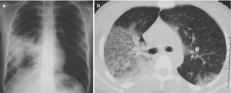
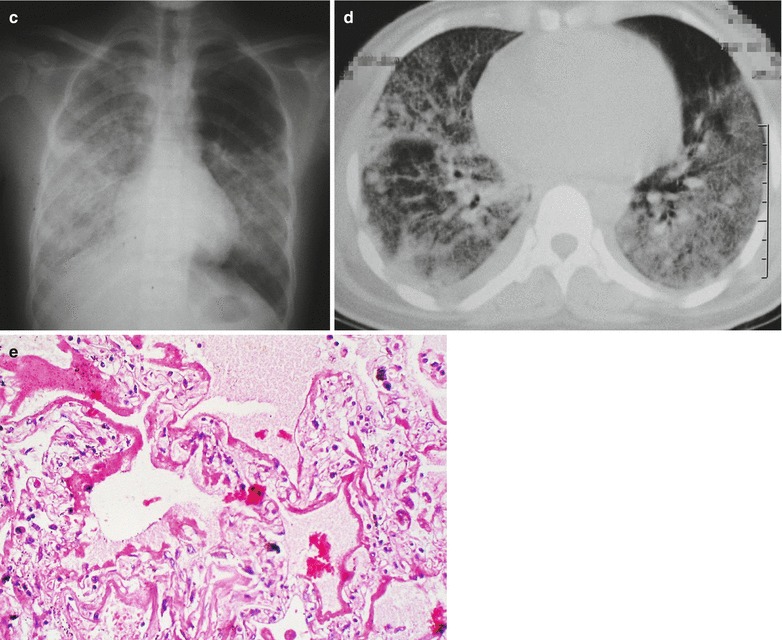
SARS and ARDS. (a) At day 9 of the illness course, chest X-ray demonstrates large flake of shadow with high density at the right lower and middle lung fields. (b) CT scanning demonstrates ground-glass shadow at the right lung and dorsal segment of the left lower lung lobe, which is more obvious at the right lung. (c) At day 11 of the illness course, chest X-ray demonstrates white lung sign at the right lung and the left lower and middle lung fields. (d) CT scanning demonstrates diffuse ground-glass shadow and consolidation at both lungs. (e) Pathology demonstrates evenly light-stained acidophilic exudated fluid filled in the alveolar cavity that is serous or cellulosic fluid and formation of hyaline membrane by condensed exudates that adheres to the alveolar wall (H&E, ×200)