
Fig. 20.6.
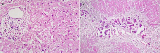
Hepatic pathological changes of SARS. (a) Swollen hepatocytes, proliferation of intralobular Kupffer cells, slightly enlarged portal area, and infiltration of small quantity monocytes (H&E, ×400). (b) Flakes and strips of necrosis of hepatocytes at lobular area III (H&E, ×400)