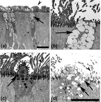
Fig. 2.

Morphological features of columnar epithelial cells in HAE. a Histological section of HAE showing ciliated and nonciliated columnar epithelial cells with distribution similar to human tracheobronchial airway epithelium in vivo. b–d Transmission electron micrographs detailing morphological characteristics of nonciliated columnar epithelial cells in HAE. b A frequently identified nonciliated columnar cell containing clear mucin-rich granules and resembling a Goblet cell. c An infrequently identified nonciliated columnar epithelial cell with electron dense granules and resembling a Serous cell. d An infrequently identified nonciliated columnar epithelial cell with mixed phenotypes of mucin-secreting and serous-like cell features. Arrowheads indicate cilia of ciliated columnar cells and long arrows indicate clear and/or dense granules in nonciliated columnar epithelial cells. Scale bars represent 20 μm
