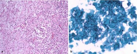
Fig. 22.15.

(a, b) A 52-year-old female with a left frontal lobe intraparenchymal abscess caused by Nocardia sp. Figure (a) shows the thickened collagenous abscess wall with neovascularization adjacent to acute inflammatory infiltrate. H&E, 10×. In figure (b), Gomori methenamine silver (GMS) stain highlights numerous branching, filamentous bacteria. GMS, 60× (Both courtesy of Anthony Yachnis, MD, and Kelly Devers, MD, University of Florida College of Medicine)
