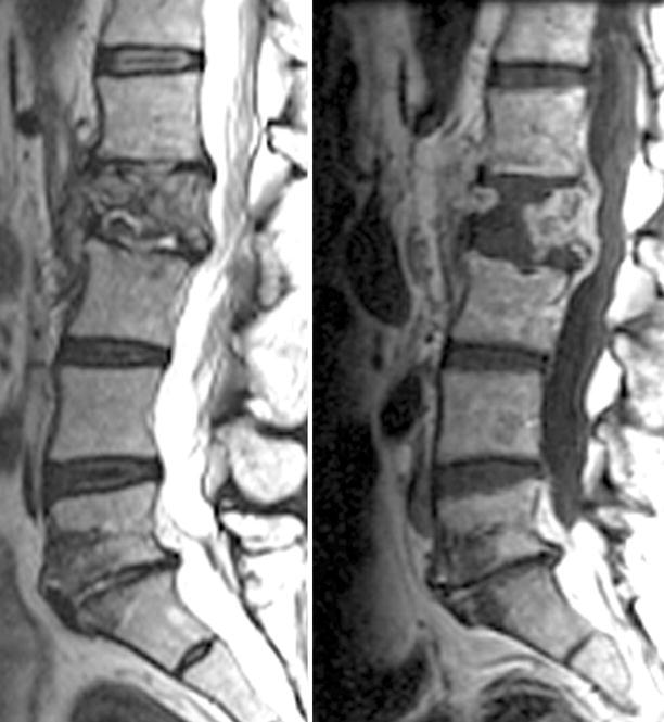
Fig. 22.18.

Spinal vertebral osteomyelitis with relative sparing of the disc space; images include T2-w sagittal image (left) and a post-contrast T1-w image (right). In this instance the spinal infection has virtually destroyed the L1 vertebral body. There is prominent residual contrast enhancement in the affected vertebral body. There is only minimal edema in the L1–2 disc space and relatively little enhancement. This complex would be consistent with hematogenous seeding the vascularized vertebral body rather than a primary disc infection with secondary osteomyelitis
