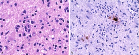
Fig. 22.27.

(a) CNS toxoplasmosis. This figure features cysts (bradyzoites) of Toxoplasma gondii in brain tissue (H&E, 60×). (b) Shows an anti-Toxoplasmagondii antibody immunohistochemical study that is immunoreactive for both bradyzoites and trophozoites (Both courtesy of Anthony Yachnis, MD, and Kelly Devers, MD, University of Florida College of Medicine)
