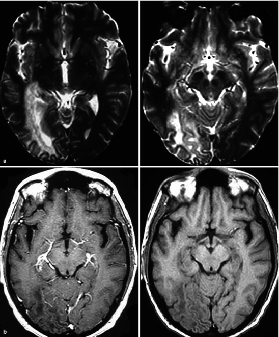
Fig. 22.30.

(a, b) Progressive multifocal leukoencephalopathy (JC virus). Figure (a) includes two adjacent T2-w sections. Figure (b) includes a post-contrast (left) and pre contrast (right) images of the same area. These images demonstrate the features of PML with a focal area of abnormality affecting mainly white matter with no appreciable internal contrast enhancement. These findings are nonspecific but in the context of an immune compromised host, PML is an important consideration. PML can cross the midline through the corpus callosum in which case it simulates both lymphoma and diffusely infiltrating astrocytoma
