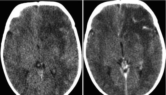
Fig. 22.8.

Acute left frontal lobe bacterial cerebritis; images include pre and post-contrast CT sections sagittal projection lower thoracic area. The early phase of brain infection (early cerebritis) demonstrates nonspecific cerebral edema and poorly defined contrast enhancement. There is frequently reactive pial hyperemia. In later stages the cerebritis will organize into early then mature stages of brain abscess
