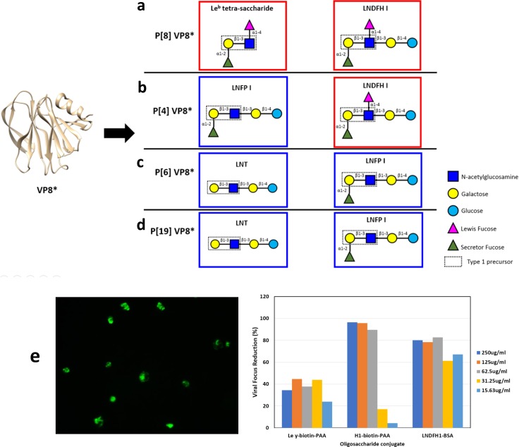
Fig 1. Schematic representation of VP8* from P[II] genogroup with its corresponding binding glycan structures.
(a) Schematic representation of P[8] VP8* and its binding glycan structures, Leb tetra-saccharide and LNDFH I. (b) Schematic representation of P[4] VP8* and its binding glycan structures, LNDFH I and LNFP I. (c) Schematic representation of P[6] VP8* and its binding glycan structures, LNT and LNFP I. (d) Schematic representation of P[19] VP8* and its binding glycan structures, LNT and LNFP I. The red boxes encircle Leb glycans and the blue boxes encircle non Leb glycans. (e) Inhibition of rotavirus Wa strain replication in HT29 cells. Left) Representative indirect immunofluorescence assay (IFA) microscopy image of P[8] RV Wa strain replication focuses in HT29 cells. Right) Quantitation of viral replication focus reductions observed in wells incubated with LNDFH1-BSA and H type 1-biotin-PAA in compared with cell culture wells with media only. Ley-PAA is a negative ligand control.

