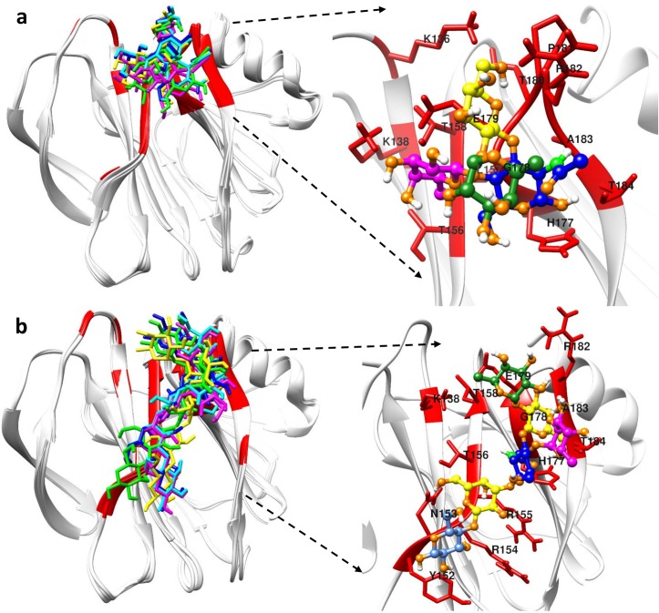
Fig 7. HADDOCK docking results of P[8] with its glycans.
(a) Cartoon figures in the left panel show the superposition of the top five best-scoring Leb tetra-saccharide bound P[8] VP8* structures. The right panel in (a) highlights the binding pocket of P[8] in recognizing Leb tetra-saccharide. (b) The superposition of the top five best-scoring LNDFH I bound P[8] VP8* structures is shown in the left panel. The right panel in (b) highlights the binding pocket of P[8] in recognizing LNDFH I. Red colors in the proteins represent the binding interface. Different colors were used to represent different carbohydrate moiety of the ligand: Lewis fucose (magenta), secretor fucose (green), galactose (yellow), N-acetylglucosamine (blue), glucose (cornflower blue).

