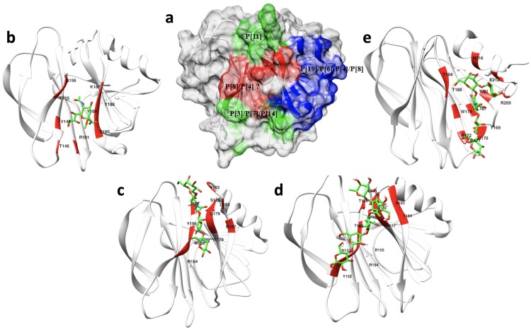
Fig 10. The binding interface of different genotypes of VP8* in recognizing their corresponding glycans.
(a) Surface-rendered structure of the apo P[6] VP8* domain [PDB ID: 6NIW] as a representative of the VP8* structures depicting a summary of the distinct glycan binding sites identified to date. The red-colored region indicates the ββ binding motif that P[3]/P[7] use in binding sialic acid, P[14] in binding type A HBGAs, P[11] in binding type 1 HBGAs, and P[8] in recognizing Lewis type HBGAs. The blue-colored region indicates the βα binding pocket that the P[19], P[6] and P[4] RVs use in recognizing H-type 1 HBGAs. (b) Detail of the sialic acid binding interface (red) of P[3] [PDB ID:1KQR]. (c) Detail of the LNT binding site of P[11] [PDB ID: 4YFZ]. (d) Detail of the LNDFH I binding interface of P[8]. (e) Detail of the LNFP I binding site of P[4] [PDB ID: 5VX5].

