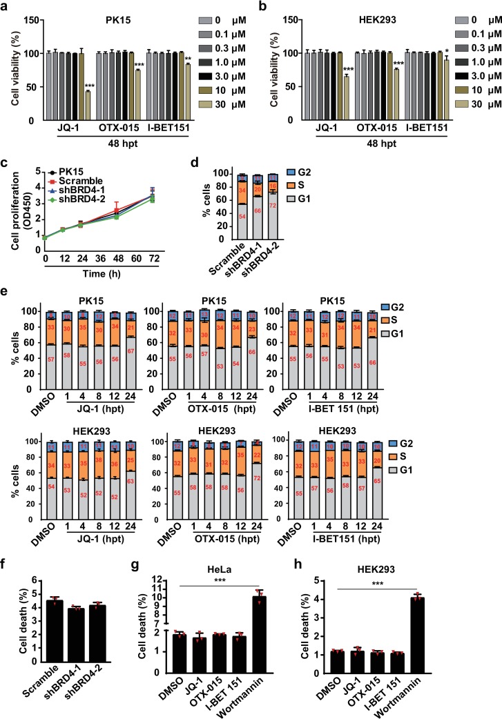
Fig 2. BRD4 inhibition induces cell cycle arrest but not apoptosis.
(a and b) Cell viability was assessed with CCK-8 cell counting assays in PK15 and HEK293 cells treated with 0–30 μM of JQ-1, OTX-015 and I-BET 151 at 48 hpt. Data are shown as mean ± SD based on three independent experiments. * P < 0.05, ** P < 0.01, *** P < 0.001 determined by two-tailed Student’s t-test. (c) Cell proliferation was assessed with CCK-8 cell counting assays in Scramble, shBRD4-1 and shBRD4-2 PK15 cells cultured for 0, 12, 24, 36, 48, 60 and 72 h. (d) The cell cycle was assessed with flow cytometry in Scramble, shBRD4-1 and shBRD4-2 PK15 cells cultured for 24 h. (e) The cell cycle was assessed with flow cytometry in PK15 and HEK293 cells treated with DMSO, JQ-1 (1 μM), OTX-015 (10 μM) and I-BET 151 (10 μM) at 1, 4, 8, 12 and 24 hpt. (f) Apoptosis was assessed with flow cytometry in Scramble, shBRD4-1 and shBRD4-2 PK15 cells cultured for 24 h. (g and h) Apoptosis was assessed with flow cytometry in HeLa (g) and HEK293 (h) cells treated with DMSO, JQ-1 (1 μM), OTX-015 (10 μM), I-BET 151 (10 μM) and wortmannin (2.5 μM) for 24 h. Data are shown as mean ± SD based on three independent experiments. *** P < 0.001 determined by two-tailed Student’s t-test.

