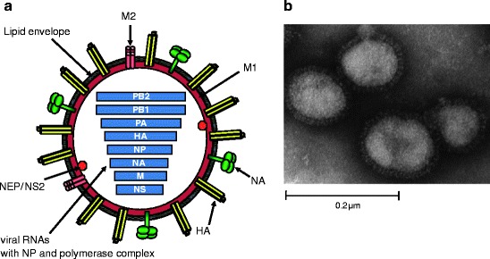
Fig.1.

Schematic structure and electron micrograph of influenza virus A. (a) The viral envelop anchors the HA and NA glycoproteins and M2 protein and is derived from the host cell during the process of budding. M1 lies beneath the viral envelope. NEP/NS1 and the core of the virion are contained within. The core consists of eight segments of viral RNA associated with the polymerase complex (PB2, PB1, and PA) and NP. Adapted from [1] and kindly provided by M.L. Shaw. (b) Negatively stained electron micrograph of mouse-adapted influenza A WSN/33. Glycoprotein spikes are visible on the surface of the virion. Kindly provided by M.L. Shaw
