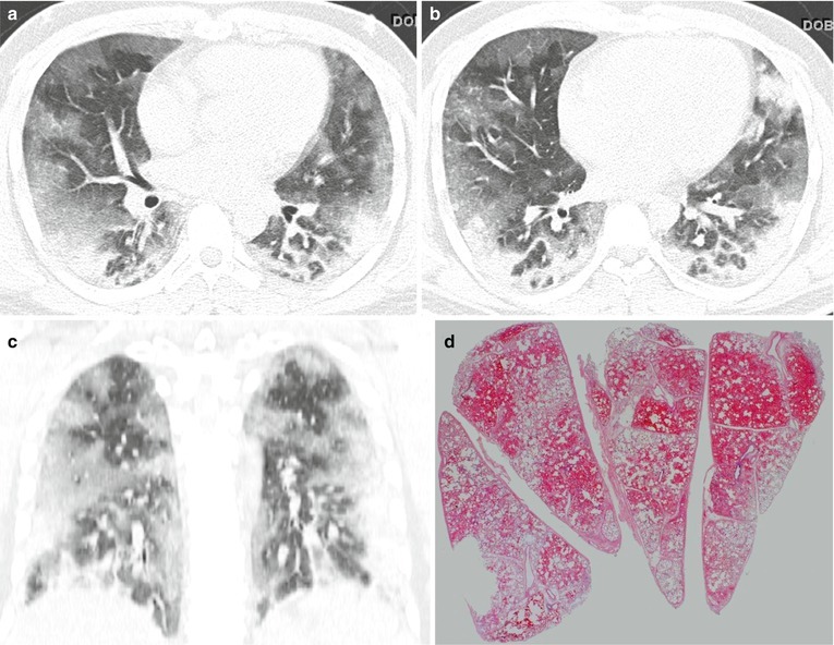
Fig. 22.8.

Diffuse alveolar hemorrhage in a 27-year-old man without specific underlying disease identified. (a, b) Lung window images of CT scans (2.5-mm section thickness) obtained at levels of right middle lobar bronchus (a) and segmental bronchi (b), respectively, show diffuse areas of parenchymal opacity (consolidation and ground-glass opacity) in both lungs. (c) Coronal reformatted image (2.0-mm section thickness) demonstrates diffuse areas of parenchymal opacity in both lungs. (d) Low-magnification (×10) photomicrograph of surgical biopsy specimen obtained from right lower lobe exhibits alveolar filling with diffuse intra-alveolar hemorrhage. Specific cause of diffuse alveolar hemorrhage was not elucidated with additional histopathologic studies
