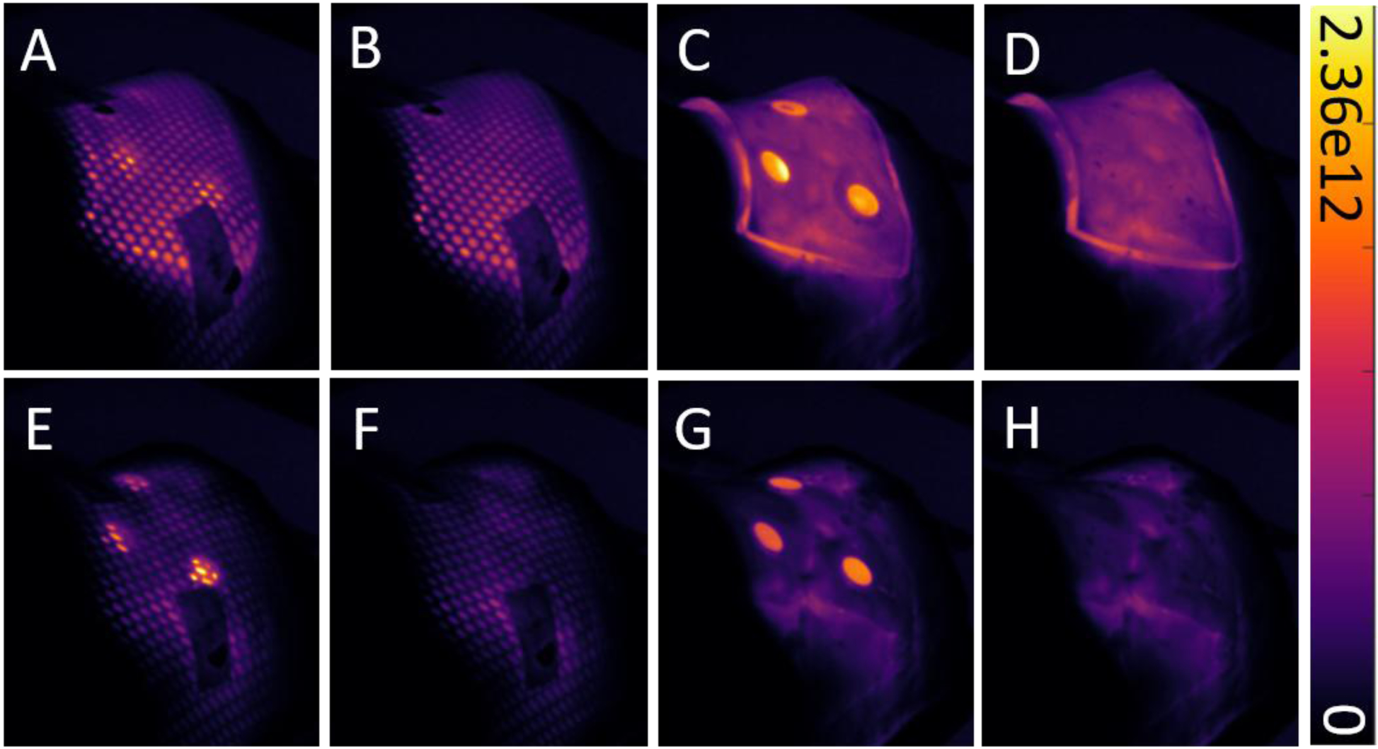
Figure 5:

Cumulative images showing various combinations of treatment materials applied to a phantom during irradiation, with and without scintillators. The same head and neck VMAT plan was administered in all eight scenarios. The top row (A-D) shows images from treatment with transparent bolus, while the bottom row (E-F) shows images from treatment without bolus. Colorbar is presented in photons cm−2 sr−1 s−1.
