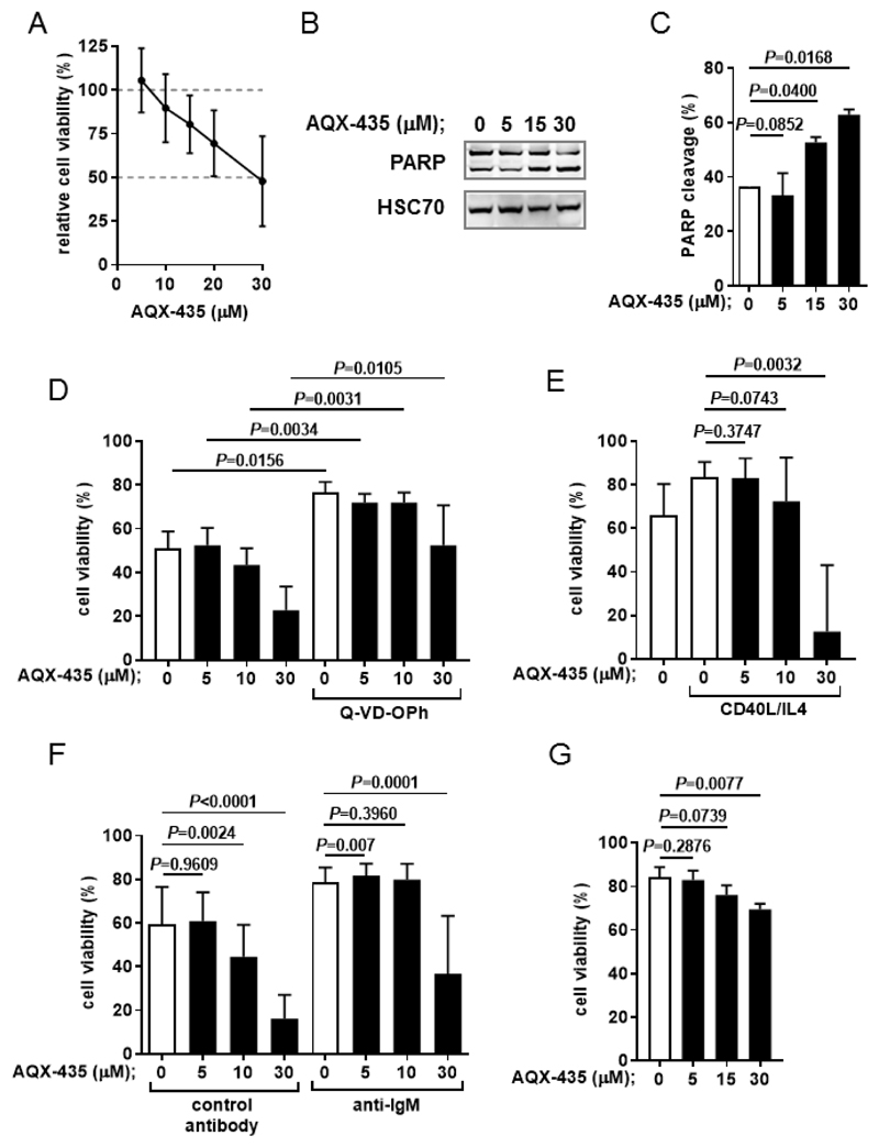
Figure 4.
AQX-435 induces caspase-dependent apoptosis of CLL cells and overcomes the survival promoting effects of microenvironment-related factors. A, CLL samples (n=24) were treated with various concentrations (5–30 μM) of AQX-435 or DMSO as a control for 24 hours and cell viability was analyzed using annexin V/PI staining. Graph shows mean (±SD) percentage of viable (annexin V-/PI-) cells. B and C, CLL samples were treated with the indicated concentrations of AQX-435 or DMSO as a control for 24 hours and PARP expression was analyzed by immunoblotting. B, Representative results and C, quantitation for all samples analyzed (n=3). D, CLL samples (n=4) were incubated with the indicated concentrations of AQX-435 or DMSO in the presence or absence of Q-VD-OPh (10 μM). After 24 hours, cell viability was analyzed using annexin V/PI staining. E and F, CLL samples were incubated with the indicated concentrations of AQX-435 or DMSO in the presence or absence of E, CD40L/IL4 (n=10) or F, bead-bound anti-IgM or control antibody (n=9). After 24 hours, cell viability was analyzed using annexin V/PI staining. G, PBMCs from healthy donors (n=5) were treated with the indicated concentrations of AQX-435 or DMSO. After 24 hours, cells were stained with anti-CD19, anti-CD5, annexin V-FITC and PI and analyzed by flow cytometry. Graphs C to G, show mean (±SD) percentage of PARP cleavage or viable (annexin V-/PI-) cells and the statistical significance of the indicated differences (Student’s t-test).

