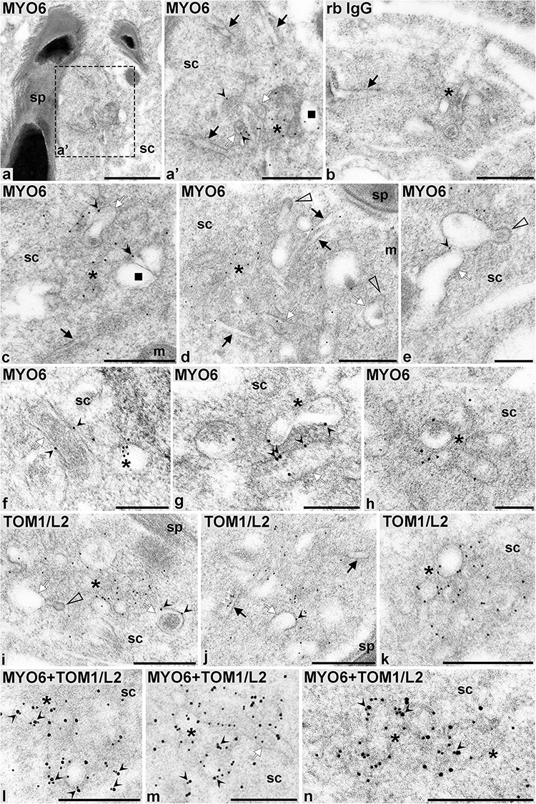
Figure 4.

MYO6 and TOM1/L2 co-localize on vesicular structures and at the bulbar region of the TBCs. Detailed immunogold localization of MYO6 (a, a’, c–h), TOM1/L2 (i–k), double-localization of MYO6 and TOM1/L2 (l–n), and negative control (b) in sv/+ spermatids. On, images l-n MYO6 was localized with 10 nm and TOM1/L2 with 15 nm gold particles. Asterisks: area enriched in endocytic structures, black arrows: proximal tubules of TBCs, black squares: larger endosomes, white arrows: bulbs of TBCs, white arrowheads: clathrin-coated pits of TBCs, sc: Sertoli cell, sp: spermatid. Bars: 1 μm (a), 500 nm (a’, b–d, i, j), 250 nm (e–h, k–n).
