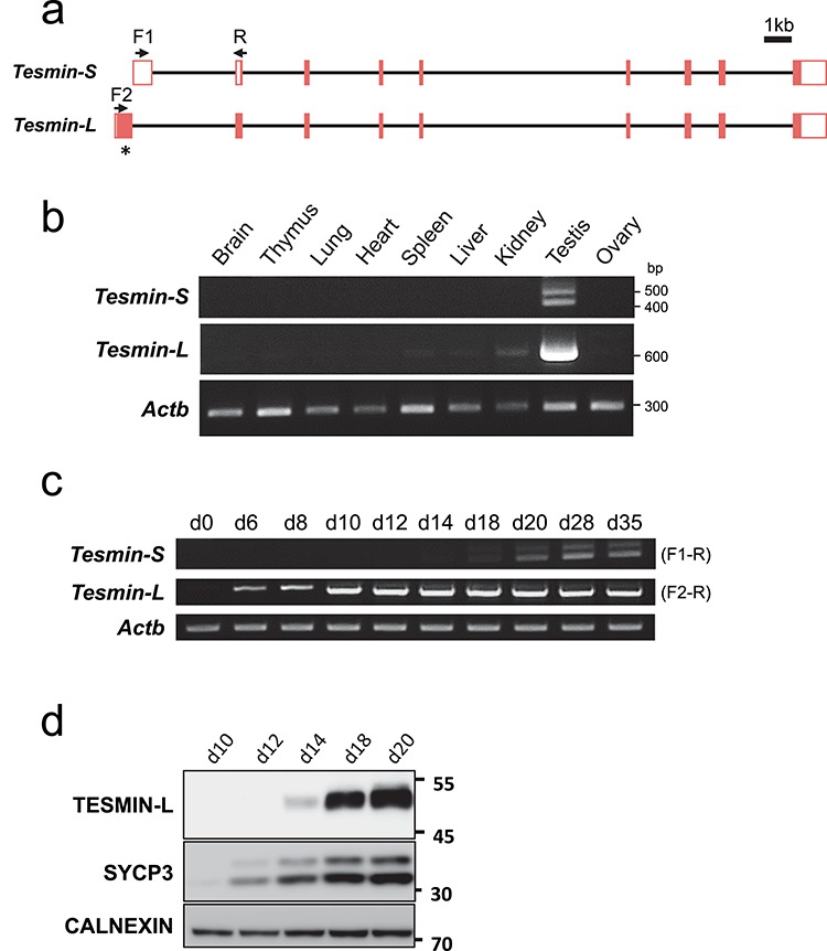
Figure 1.

Expression profile of Tesmin isoforms. (a) Schematic representation of two splicing variants of mouse Tesmin. Filled box: protein-coding sequence, white box: untranslated sequences, arrows: primer binding sites. * shows the region that the antibody was raised against. (b) RT-PCR of Tesmin using cDNAs obtained from various tissues. Actb as control. (c) Expression of each variant analyzed with RT-PCR using testicular cDNA obtained from 0-, 6-, 8-, 10-, 12-, 14-, 18-, 20-, 28-, and 35-day-old mice. Actb as control. Primer sets used for each Tesmin-S and Tesmin-L are indicated at right, whose annealing sites are shown in Figure 1a. (d) TESMIN-L expression in spermatogenesis detected by western blotting. SYCP3 (a meiosis marker) and CALNEXIN (ubiquitously expressed marker) were used as controls.
