Figure 3.

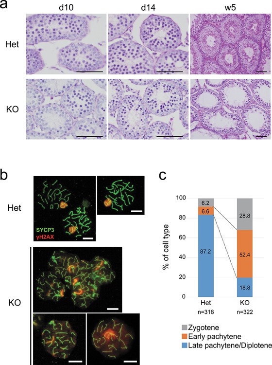
Spermatogenesis arrest occurred at meiosis. (a) PAS–hematoxylin staining of the testis from Tesmin +/em1 and 10-, 14-day-old, and 5-week-old KO mice . Scale bars: 50 μm. (b) Immunostaining of meiotic spreads from Tesmin +/em1 and KO spermatocytes for the synaptonemal complex component SYCP3 (green) to identify chromosome axes, and γH2AX (red) as a marker for DNA damage. Three spermatocytes from heterozygous are in late-pachytene stage. Top and bottom left spermatocytes of KO are categorized as early pachytene stage, and bottom right is categorized as late pachytene stage. Scale bars: 10 μm. (c) Percentage of spermatocytes from Tesmin +/em1 and KO male mice that are categorized as late zygotene, early pachytene and late pachytene by the feature of SYCP3 and γH2AX staining. Three mice (over 5 weeks old) are used for each genotype and more than 50 nuclei were observed from one mouse. The numbers in bar graph indicate the ratio (%) of each category. P-values of each categories are; zygotene: 0.0373, early pachytene: 0.0033, and late pachytene/diplotene: 0.0002, respectively. Welch t test was used for statistical analysis.
