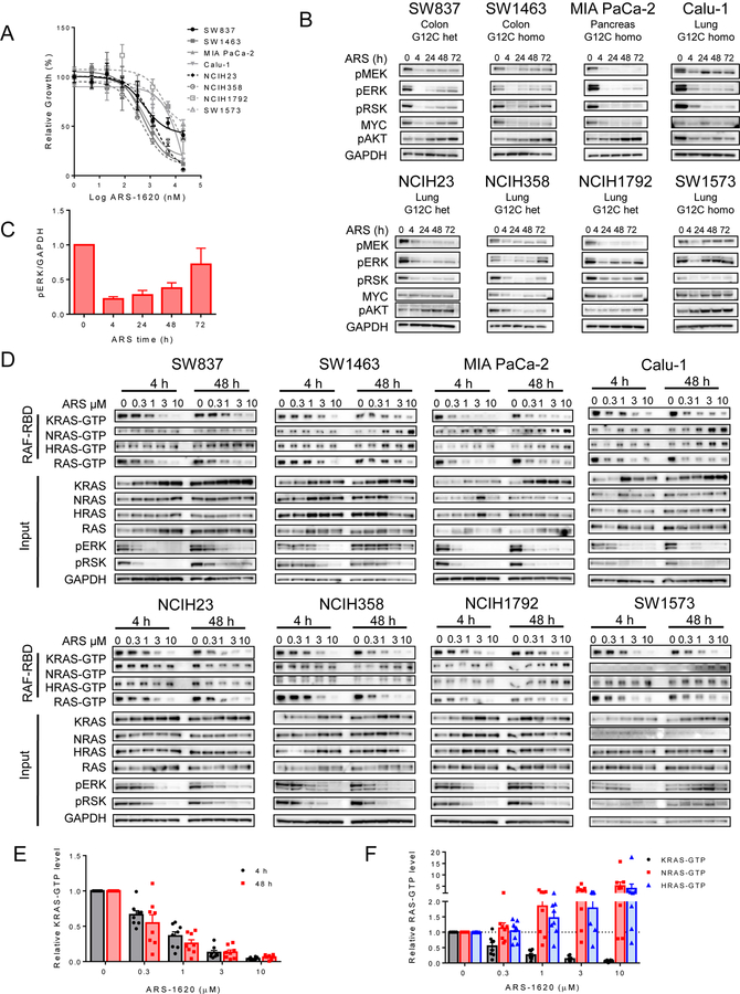
Figure 1. Feedback reactivation of RAS signaling occus following KRASG12C inhibition.
(A) Cell lines were treated for 72 h with a dose titration of ARS-1620 and viability was measured by CellTiter-Glo. (B) KRAS-G12C mutant cell lines were treated with ARS-1620 (1 µM) for 0, 4, 24, 48, and 72 h. Blot analysis was performed for phospho- (p)MEK, pERK, pRSK, pAKT, and total MYC with GAPDH as a loading control. (C) Densitometry of phospho-ERK normalized to GAPDH for blots in, results represent an average of phospho-ERK across all 8 cell lines (A).(D) Cell lines were treated with a dose titration of 0.3–10 µM ARS-1620 for 4 or 48 h and lysates were subject to a RAF-RBD pulldown and blot analysis of KRAS, NRAS, HRAS and total RAS as well as pERK, pRSK and GAPDH for input samples. (E) Densitometry analysis of 4 and 48 h KRAS-GTP levels normalized to input KRAS and GAPDH loading control in D (F) Densitometry analysis of 48 h KRAS-GTP, NRAS-GTP, and HRAS-GTP levels normalized to input RAS and GAPDH loading control of blots in (D). Densitometry results in (E) and (F) represent an average across all 8 cell lines.

