
Fig 1. Folate supplementation of Sprague-Dawley rat progenitors enhances in vivo regeneration of injured CNS axons into a peripheral nerve graft in untreated transgenerational progeny.
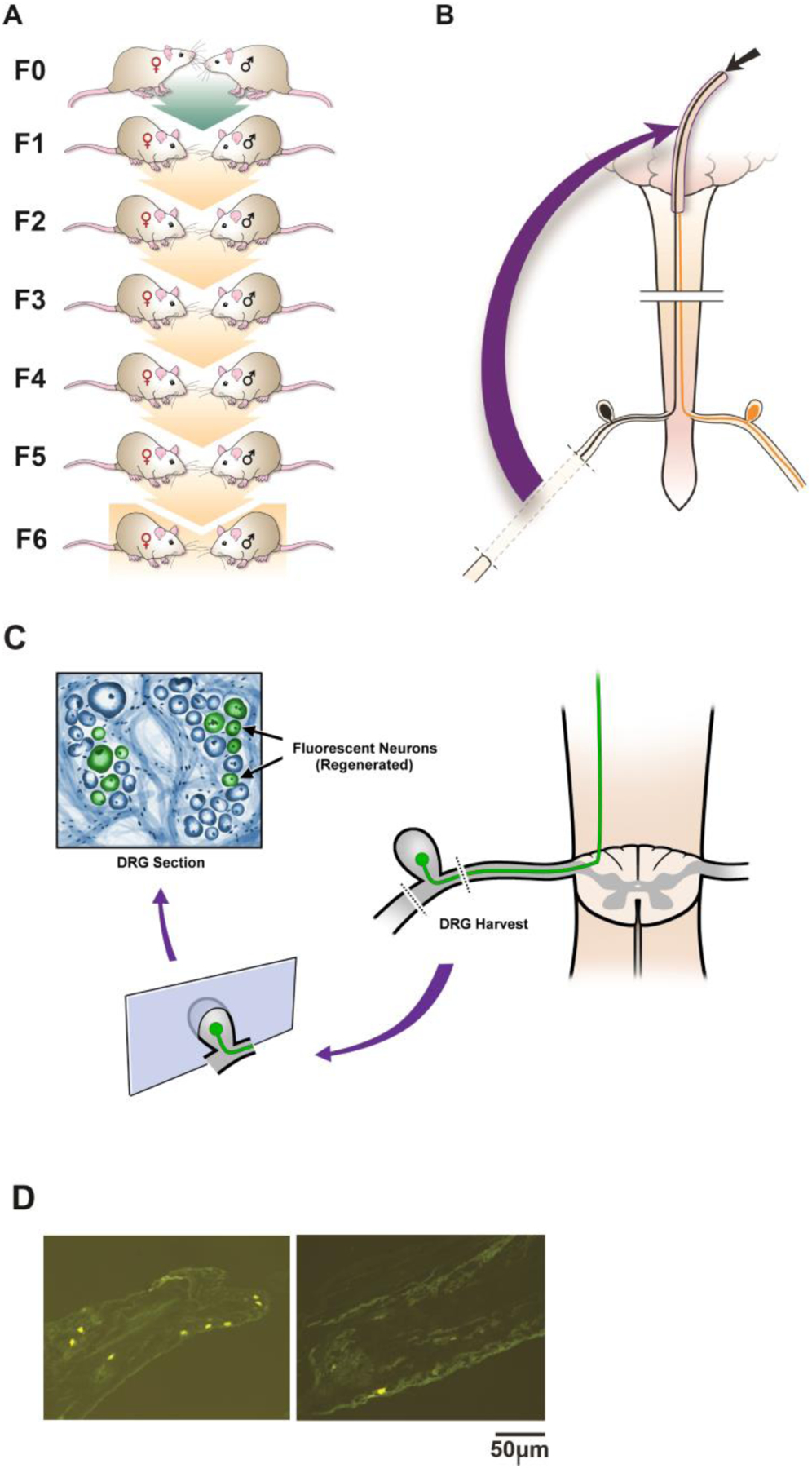
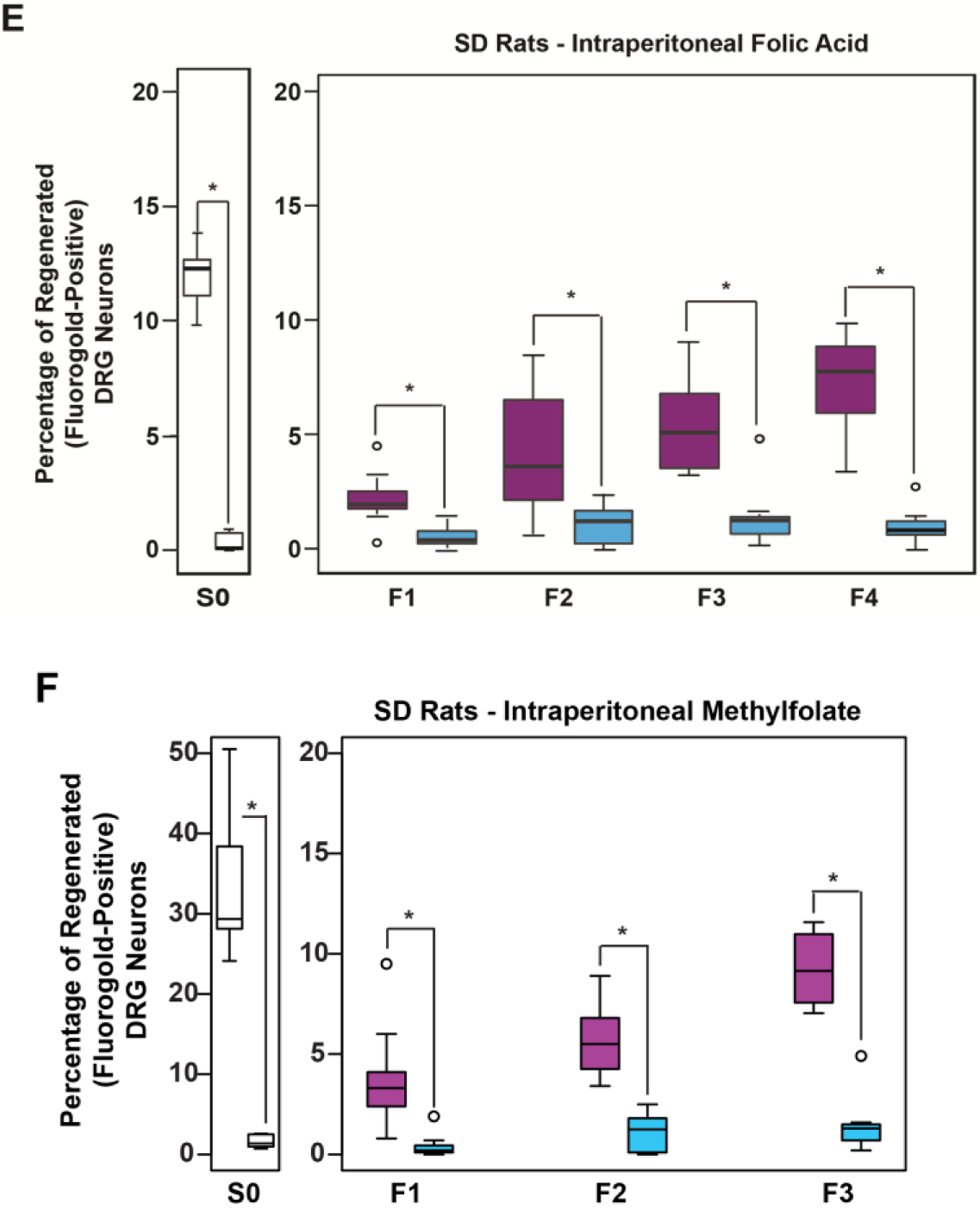
(A-C) Rat Spinal Cord Regeneration Model(SCRM): Experimental Design Schematic: (A) Mating pairs of animals (F0) were treated with folate or vehicle starting 2 weeks before breeding, and continuing in females until weaning, and in males until pregnancy was assured. Four to six generations of progeny (depending on intervention) were bred without treatment. (B) A sciatic nerve graft is implanted at the site of bilateral C3 dorsal column lesion (purple arrow). At 2 weeks, a fluorescent tracer is placed at the free end of the graft (black arrow), and (C,D) its uptake is detected 48 hours later in the lumbar DRG neuron cell bodies of axons that have extended into the graft ipsilateral to sciatic nerve harvest. The fluorescent tracer is taken up only by regenerated neurons. (E) Intraperitoneal (IP) folic acid supplementation of progenitors enhances spinal axon regeneration in untreated F1-F4 male progeny. Percentage of DRG neurons of the untreated offspring (F1-F4) of treated progenitors that have regenerated spinal axons into sciatic nerve grafts compared to DDI controls. Single generation control animals (S0) with direct exposure to folate supplementation exhibit approximately 12% regeneration. Animals whose progenitors (F0) were supplemented with folate regenerate injured spinal axons for 4 generations (n: (FA-purple, DDI-blue) = S0: 8, 8; F1: 12, 8; F2: 16, 10; F3: 10, 8; F4: 10, 10; *p<0.05). (F) Intraperitoneal methylfolate supplementation of progenitors enhances spinal axon regeneration in untreated F1-F3 progeny. Percentage of DRG neurons of the untreated offspring (F1-F3) of treated progenitors that have regenerated spinal axons into the sciatic nerve grafts compared to DDI controls (n (MF - purple, DDI- blue) = S0: 12, 7; F1: 14, 11; F2: 12, 10; F3: 8, 8; *p<0.05).