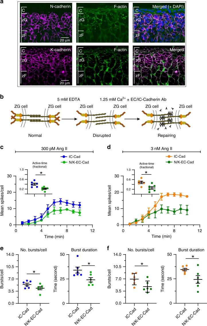
Fig. 7. Disruption of N-cadherin shortens burst duration without affecting other calcium spike parameters.
a Immunohistochemistry showing N-cadherin (red, upper left), K-cadherin (red, lower left) and F-actin (green, middle) are abundant in the zG layer (right, merged images). Scale bar: 20 µm. b Model of calcium switch assay: Disrupting adherins junctional complex by reducing extracellular calcium with EDTA. Anti-N/K-cadherin antibodies (EC-Abs) binds to the now exposed extracellular N- and K-cadherin binding sites, inhibiting reformation of strong cell–cell adhesion after calcium is restored. For controls, we substituted an antibody directed to the conserved intracellular domain (IC-Ab) of cadherins. c–f Adrenal slices were stimulated with 300 pM (c, e) or 3 nM (d, f) Ang II after 1.5 min of baseline recording. EC-Abs: n = 6; IC-Ab: n = 6. c, d Mean spikes per 1 min bin over a 10 min period; EC-Abs treated slices had fewer spikes over time for both 300 pM and 3 nM Ang II (2-way ANOVA, effect of antibody: P = 0.011 and 0.018, respectively) as well as mean fractional active time (c, d insets, mean ± SEM: 300 pM: IC-Ab 0.40 ± 0.04, EC-Abs 0.22 ± 0.03; 3 nM: IC-Ab 0.48 ± 0.05, EC-Abs 0.26 ± 0.06; Wilcoxon rank test, *P = 0.016 and 0.016, respectively). e, f Reduced activity was due to a decrease in the number of bursts per cell (e, f left panels; mean ± SEM: 300 pM: IC-Ab 5.43 ± 0.39, EC-Abs 4.35 ± 0.44; 3 nM: IC-Ab 6.94 ± 0.67, EC-Abs 4.8 ± 0.66; Wicoxon rank test; 300 pM: *P = 0.047, 3 nM: *P = 0.016) and a shortening of burst duration (e, f right panels; mean ± SEM: 300 pM: IC-Ab 34.1 ± 3.10, EC-Abs 24.2 ± 2.25; 3 nM: IC-Ab 34.54 ± 1.36, EC-Abs 24.38 ± 3.94; Wilcoxon rank test; 300 pM: *P = 0.016, 3 nM: *P = 0.047). Mean data from each mouse were represented as a single point in calculating N/experimental condition. c, d Lines and symbols are color coded according to experimental condition; IC-Ab + 300 pM Ang II: blue, EC-Abs + 300 pM Ang II: light green, IC-Ab+ 3 nM Ang II: orange, EC-Abs + 3 nM Ang II: dark green. Source data are provided as a Source Data file.

