Figure 1.
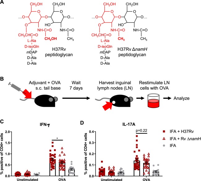
CFA-dependent cell-mediated immune responses as a function of mycobacterial namH. (A) PGN of wild-type H37Rv M. tuberculosis (left) and PGN of the ΔnamH mutant (right). The MDP motif is drawn in red, and the site of N-glcolylation is in bold font. With NamH, N-glycolylation was shown on ~70% of muramic acid residues, with N-acetylation on the remaining ~30%8,11. (B) Immunization scheme (relevant to Figs. 1, 2 and 4): mice were immunized with adjuvant emulsion containing OVA by s.c. injection at the base of the tail, and after seven days, inguinal (draining) lymph nodes were harvested. Lymph node cells were cultured ex vivo with or without OVA to examine the OVA-specific cytokine response by flow cytometry or ELISpot. (C,D) Proportion of cytokine-producing CD4+ CD8− lymph node cells of mice immunized against OVA with heat-killed M. tuberculosis strain H37Rv, H37Rv ΔnamH, or IFA alone, seven days prior. Shown are data pooled from four separate experiments with averages +/− SEM. p-values were calculated with two-tailed student’s t-tests. *p < 0.05. For IFA + H37Rv, IFA + H37Rv ΔnamH, and IFA alone, sample size N = 31, 27 and 16 mice, respectively. Each plotted point represents the result obtained from an individual mouse.

