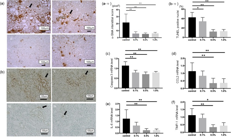
Figure 4.
The anti-apoptotic and anti-inflammatory effects of Si-based agent on renal function. (a) Interstitial phenotypic changes are assessed by immunohistochemical staining of α-SMA (arrow) in the control group (i) or the Si-based agent (0.1wt.%-1.0wt.%) groups (ii-iv). The positive area was quantitatively assessed using a color image analyzer (v). (b) TUNEL method was performed for measuring the levels of apoptosis in each group as describe above (i-iv). We counted the number of TUNEL-positive cells (arrow) per 100 cells in ten random areas of each rats (v). Scale bar: 100 µm. (c-f) The normalized messenger RNA (mRNA) level of caspase-3 (c), C-C motif chemokine ligand 2 (CCL2) (d), interleukin-6 (IL-6) (e), and a tissue inhibitor of metalloproteinases (TIMP-1, f) were measured. The levels are normalized to β-actin levels for each sample. The y-axis values represent the number of copies relative the number of copies in the same samples. The levels are normalized to β-actin levels for each sample. The y-axis values represent the number of copies relative the number of copies in the same samples. Data were expressed as mean +/− SD. **p < 0.01, *p < 0.05 vs. the control group, respectively.

