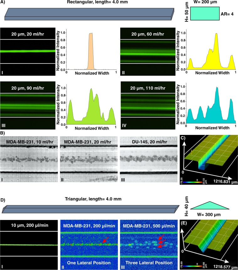
Figure 4.
(A) Inertial microfluidics in a rectangular straight microchannel with height and width of 50 µm and 200 µm, respectively. I. At first, 20 µm particles occupy the center of the channel as their focusing position. The intensity profile also illustrates that particles are focused at the center of the channel. II–IV. Later, looking at lateral position and intensity profiles reveal that by increasing the flow rate, side walls are added to the focusing position of the particles, and the focusing band of particles at center becomes wider. To extract these images, we have used “max intensity” feature from Fiji Software (https://fiji.sc). (B) The equilibrium position of MDA-MB-231 cells at flow rates of I. 10 ml/hr and II. 20 ml/hr and III. DU-145 cells at flow rate of 20 ml/hr confirms the single-line focusing of cells within the rectangular straight microchannel (from top view). (C) A surface profilometry of the rectangular cross-section which shows the rectangular profile of the microchannel. Results show that the channel has perfect shape and quality which is suitable for inertial microfluidics. (D) In triangular microchannel, particles first migrate to I. and II. one focusing position and then this increases to III. three separate points. This trend is similar to those reported in the literature14,53,54. The results for MDA-MB-231 cells at flow rate of 200 µl/min illustrate a single-line focusing position, and at flow rate of 500 µl/min depict three focusing positions. (E) Surface profilometry of the triangular straight microchannel with height and width of 40 µm and 300 µm, respectively.

