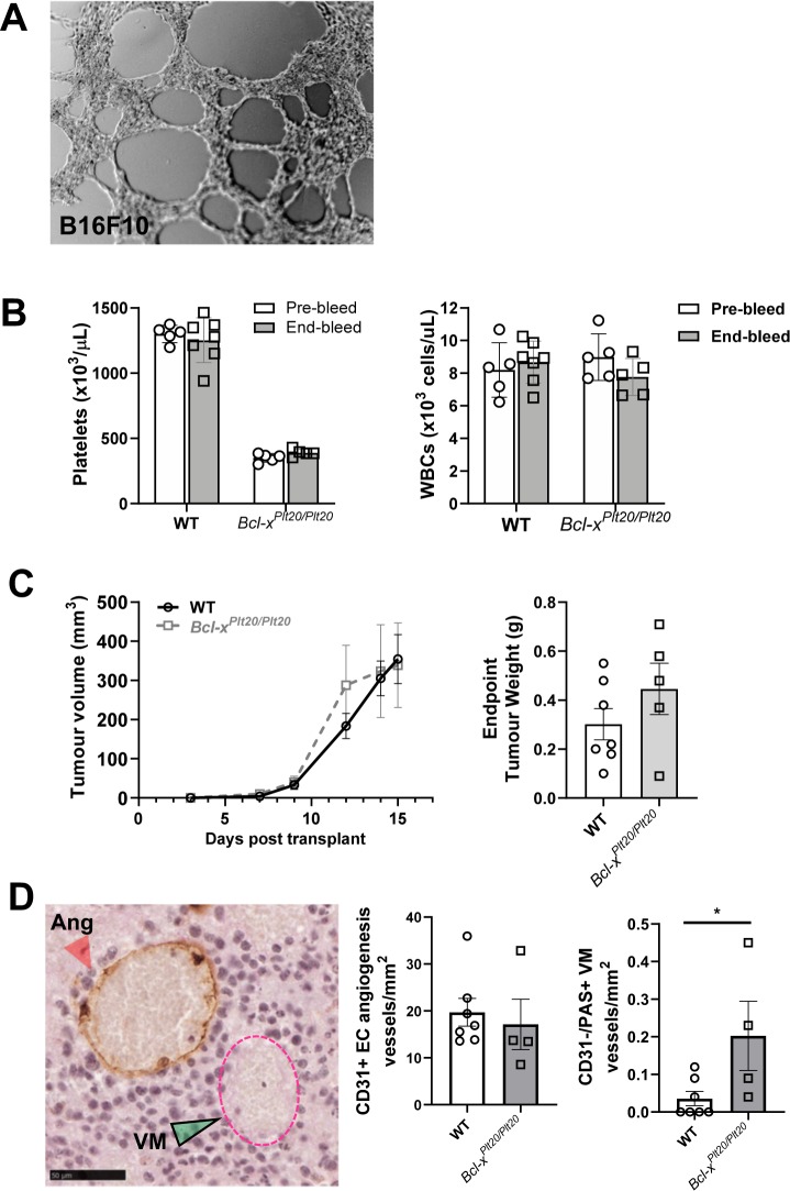
Figure 4.
VM formation by B16F10 melanoma cells and influence of platelets in vivo. In (A); representative image of B16F10 melanoma cancer cells undergoing VM in vitro in Matrigel. In (B), circulating platelet and WBC counts in wildtype (WT) and Bcl-xPlt20/Plt20 mice prior to, and experimental end (open bars, pre-bleed at day -14, grey bars, end-bleed at day 15). In (C), caliper measurements of B16F10 tumour growth over time and final B16F10 tumour weights at experimental end (open symbols, WT mice; grey symbols, Bcl-xPlt20/Plt20 mice). In (D), representative image of CD31 and PAS stained B16F10 harvested tumour. CD31+/PAS+ EC-lined angiogenic structure (Ang, red arrow head) and CD31−/PAS+ VM structure (VM, green arrow head and pink dotted line). Scale bar is 50 µm. Corresponding quantification of the average angiogenic and VM structures per mm2 (open bars, WT mice; grey bars, Bcl-xPlt20/Plt20 mice). Data show mean ± SEM for n = 5–7 mice. *p < 0.05, unpaired t-test.

