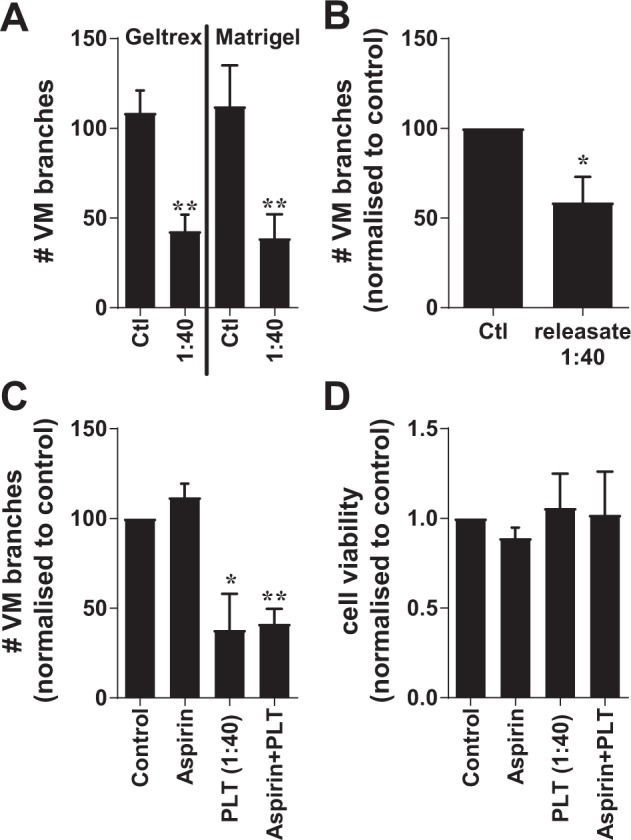
Figure 5.

VM formation and survival assays with MDA-MB-231 cancer cells in the presence of platelets, platelet releasates or Aspirin. In (A); MDA-MB-231 breast cancer cells undergoing VM in Geltrex or Matrigel in the presence of buffer control (Ctl) or platelets at the indicated ratio (cells:platelets). VM structures are expressed as mean ± SEM for n = 4–5 experiments. **p < 0.01 compared with buffer control, paired t-test. In (B); MDA-MB-231 breast cancer cells undergoing VM without and with co-culture of α-thrombin-activated platelet releasate at the indicated ratio (1:40 releasate equivalent). Data are expressed as mean ± SEM from n = 3 experiments. *p < 0.05, paired t-test. In (C), MDA-MB-231 breast cancer cells co-cultured with Aspirin (100 μM), platelets at the indicated ratio (cells:platelets) or platelets pre-treated with Aspirin (100 μM) for 10 min prior to inclusion in the VM assay. VM structures are expressed as mean ± SEM for n = 6 experiments. *p < 0.05, **p < 0.01, one-way ANOVA. In (D), MDA-MB-231 cells cultured without or with Aspirin (100 μM), platelets (1:40 ratio) or Aspirin (100 μM) pre-treated platelets prior to cell viability being examined at 24 hours via alamarBlue. Results are expressed as mean ± SEM for n = 7 experiments.
