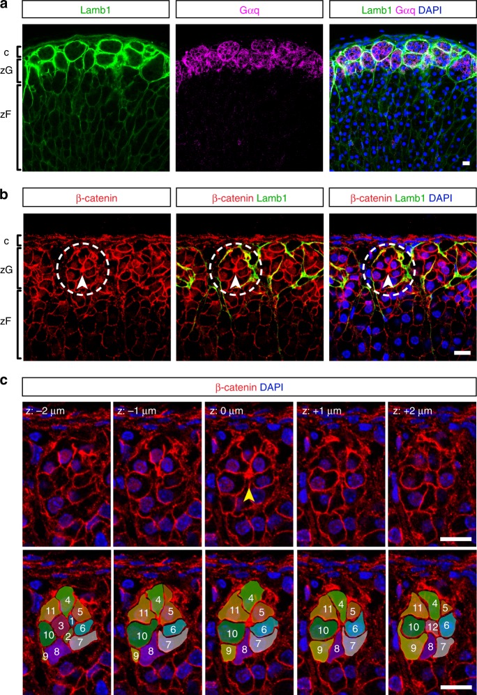
Fig. 1. Adult adrenal glomeruli consist of multicellular rosettes.
a Laminin β1 (Lamb1, green) marks the basement membrane surrounding distinct clusters of zG cells (Gαq+, magenta), defining the outline of individual glomerulus. b Cells within each glomerulus organize into rosettes. Representative image of an adult adrenal slice stained for Laminin β1 (Lamb1, green) and β-catenin (red). Dashed circles highlight a rosette example. Arrowheads point to rosette center. c Top, confocal z-stack images of the rosette encircled in (b), showing β-catenin (red) and nuclei (DAPI, blue). Z step size is 1 μm. Arrowhead points to the rosette center. Bottom, tracing of cells (pseudo-colored and numbered 1–12) participating in the rosette shown in top panel. c capsule, zG zona glomerulosa, zF zona fasciculata. DAPI (blue) marks nuclei. All bars, 10 μm.

