
Fig. 4.
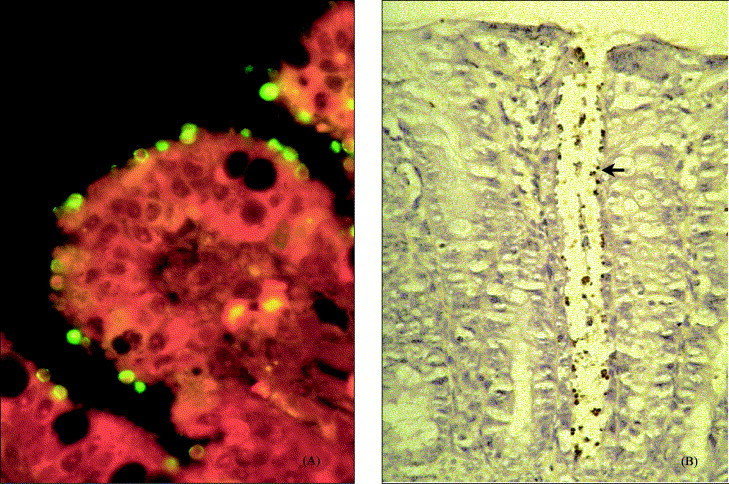
Histopathological slides (7 days post-infection) from a 9-day-old piglet experimentally infected with C. parvum (CPB-0 isolate). (A) Ileum mucosa showing stunting of villi, and numerous Cryptosporidium stages covering the surface (immunofluroescens staining; original magnification: 400×). (B) Colonic mucosa showing Cryptosporidium stages (arrow) confined to the crypt (immunohistochemistry; original magnification: 200×).