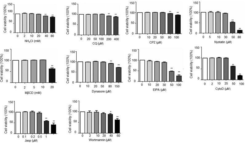
Fig. S1.
Cell viability of all the inhibitors were assessed by using the cell counting kit-8 (CCK-8). Cells were either untreated (as a control) or treated for 12 h at 37 °C with different concentrations of inhibitors. After treatment, cells were incubated with 10 μl CCK-8 solution for 2 h, and cell viability was determined by the optical density at 450 nm. The bars indicate the mean ± SD from three independent experiments. *P < 0.05; **P < 0.01 compared to the mock-treated cells.

