
Fig. 7.
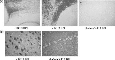
(a) In situ hybridization to localize the sites of infection by rBC, rLaSota or rLaSota V.F. virus. Three-week-old chickens were infected intranasally with 103 PFU of virus. At 1, 3, 5 and 7 days post-infection, chickens were sacrificed and perfused with 4% paraformaldehyde. Brains were divided sagitally down the midline, embedded in paraffin, and sectioned at 5 μm/section. Sections were subjected to in situ hybridization with 35S-labeled riboprobe specific for NP gene of NDV or P gene of bovine RSV. No specific signals were observed with either mock-infected chickens or with bovine RSV probe. Shown are representative sections of the cerebellum infected with rBC at 5 days post-infection (D P.I.) (A), and 7 D P.I. (B), and of cerebellum infected with LaSota V.F. virus at 7 D P.I. (C). (b) Immunohistochemistry of the brains of chicks infected with rBC, rLaSota or rLaSota V.F. virus. Three-week-old chickens were infected intranasally with 103 PFU of virus. At 1, 3, 5 and 7 days post-infection, chickens were sacrificed and perfused with 4% paraformaldehyde. Brains were divided sagitally down the midline, embedded in paraffin, and sectioned at 5 μm/section. Presence of NDV antigens was detected with a cocktail of monoclonal antibodies to NDV HN protein. (A) rBC virus infected brain shows plaque-like areas of viral antigen staining in the cerebellum at 7 D P.I. (B) rLaSota V.F. virus-infected brain shows no viral antigen in the cerebellum at 7 D P.I.