
Fig. 1.
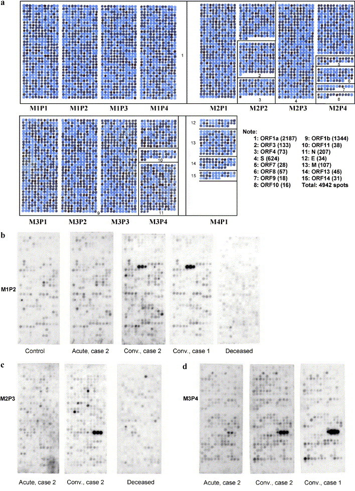
(a) Outline of the overlapping peptide set covering membranes M1, M2, M3, and M4. Panels on the membranes are designated from left to right P1, P2, P3, and P4. Numbers in the boxes designate the location of each open reading frame. The note to the right of M4P1 shows the key to the Orfs with peptide totals in brackets. This membrane was spotted with dye instead of amino acids. In actual membranes, each spot is a 10-amino-acid peptide with adjacent spots being shifted by two amino acids. Characterization of the immune response against these single case epitopes promises to provide important insights into their role in the resolution of infection. However, epitopes recognized by multiple convalescent sera may be the most important targets of neutralizing antibodies. (b–d) Examples of membrane panels probed with various serum samples and developed with peroxidase-labeled goat antihuman IgA; (b) M1P2 probed with control serum, acute as well as convalescent serum from case 2, convalescent serum from case 1, and serum from the deceased case. Notice the triad of spots recognized only in the serum of the two convalescent cases. The peptide sequences from Orf 1a are SDDYIKLNGP, DYIKLNGPLT, and IKLNGPLTVG. (c) P3M2 probed with acute and chronic serum from case 2 and serum from the deceased case. Panel 3 has peptides from S-protein. Notice the triad of spots recognized only by the convalescent serum. The peptide sequences from S-protein are FQPFQQFGRD, PFQQFGRDVS, and QQFGRDVSDF. (d) P4M3 probed with acute and convalescent serum from case 2 and convalescent serum from case 1. Notice the triad of spots recognized in the two convalescent sera. The peptide sequences from N-protein are QLPQGTTLPK, PQGTTLPKGF, and GTTLPKGFYA.