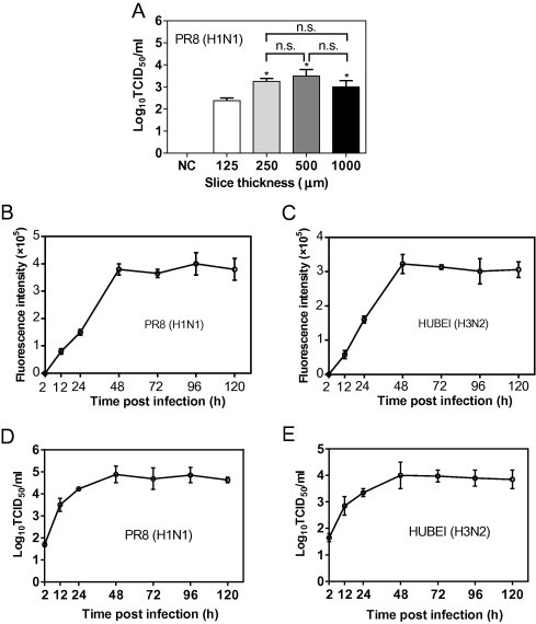
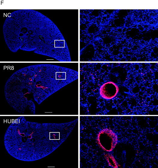
Fig. 2.


Mouse lung slices support influenza virus replication. (A) To determine the virus production of lung slices of different thickness, 125-, 250-, 500- and 1000-μm thick lung slices were exposed to 200 μl of 105 PFU/ml of PR8 (H1N1) for 48 h, and the supernatants were used to determine the virus titre by a TCID50 assay. Comparisons between groups were performed by an unpaired t-test. Data are expressed as the means ± SEM. n.s., non-significant. *, 0.01 < p < 0.05 in comparison to 125-μm thick slices. (B–E) To determine the virus yields at different time points post infection, 250-μm thick slices were infected with 200 μl of 105 PFU/ml PR8 (H1N1) (B, D) or HUBEI (H3N2) (C, E) virus. The supernatants were collected at indicated time points to perform the NA activity assay (B and C) and TCID50 assay (D and E). (F) Infection of lung slices was assessed by the IFA assay. Slices with a thickness of 150 μm were exposed to 200 μl of 106 PFU/ml PR8 (H1N1) or HUBEI (H3N2) viruses for 24 h. The slices were fixed with 4% paraformaldehyde and analysed by IFA. The images were captured using a Nikon multiphoton Confocal Microscope A1 MP+. The virus diluent was used as a negative control (NC) in the infection. Scale bar = 1000 μm. The right panels show enlarged regions of interest from the left panels.
