Figure 1.
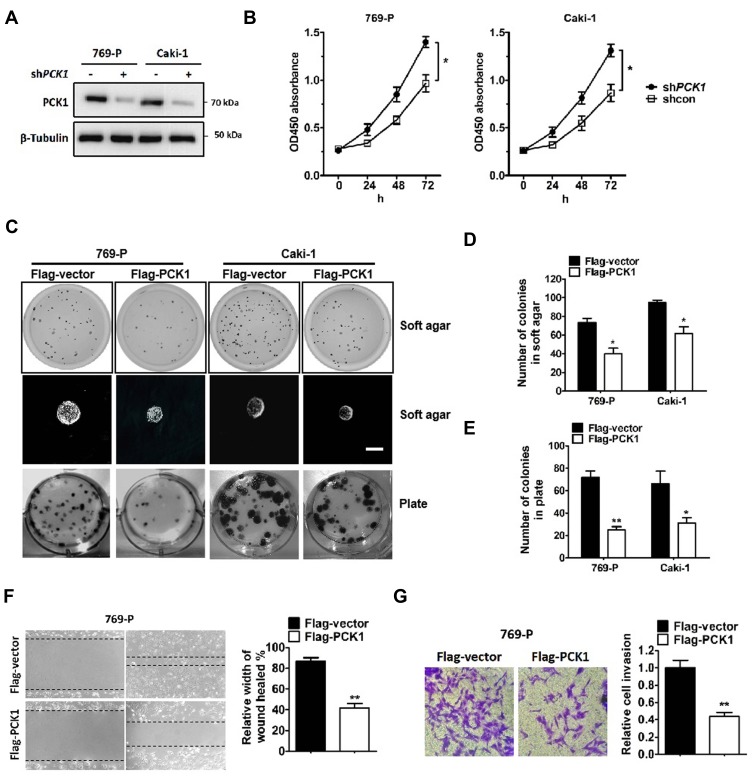
PCK1 inhibits ccRCC cell growth, colony formation, migration and invasion in vitro.
Notes: (A) 769-P and Caki-1 cells were infected with lentiviruses carrying shRNA against PCK1 (shPCK1) or negative control shRNA (shcon). PCK1 and β-tubulin levels were determined by Western blotting. (B) Cell viability was assessed by CCK-8 assays in 769-P and Caki-1 cells with stably knocked down PCK1 or the negative control. The bars represent the means±SEM (n=3). (C–E) Representative images of cell growth in soft agar colony forming assays (C, top and middle and D) and plate colony forming assays (C, bottom, and E). The bars represent the means±SEM (n=3). (F) Wound healing assay for 769-P cells stably expressing Flag-PCK1 or the control vector. The bars represent the means±SEM (n=3). (G) Transwell invasion assay for 769-P cells stably expressing Flag-PCK1 or the control vector. The bars represent the means±SEM (n=3). (*P<0.05, **P<0.01, Student’s t-test).

