
Fig. 4.
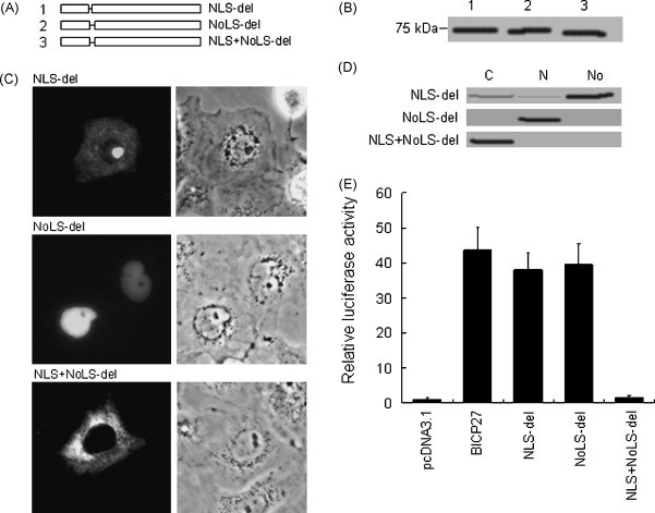
Mutation of arginine-rich amino acids residues abrogates the nuclear or nucleolar localization of BICP27. (A) Schematic diagram of deletion of arginine-rich domain in BICP27. (B) Western blotting analysis of the different deletion mutants of arginine-rich domain using EYFP polyclonal antibodies. The lane number corresponds to the constructs in Fig. 4A. (C) Subcellular localization of BICP27 arginine-rich domain deletion mutants fused with EYFP. Representative fluorescence images of the vast majority living cells expressing indicated EYFP fusion proteins and EYFP fluorescence was analyzed in living cells 24 h after transfection. (D) Western blotting analysis of the subcellular localization of different BICP27 deletions using EYFP polyclonal antibodies. (E) Transactivation analysis of gC promoter by BICP27 and its deletion mutants. Cells were harvested 48 h post-transfection and assayed for luciferase activity. Relative fold induction is calculated as light units of the test sample divided by the pGL-3 transfected cells. Standard deviations from the mean of three independent experiments are indicated.