
Fig. 9.
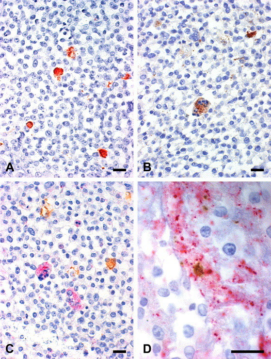
Immunohistochemical labeling of chlamydial inclusions (A, C), ca-PEDV single and syncytial cells (B) as well as viral syncytial cell with chlamydial inclusion (D). Vero cells were dually infected with C. trachomatis S45 and ca-PEDV and pelleted. Paraffin sections. Immunoperoxidase stain. (A) Chlamydial inclusions; red (AEC). (B) Ca-PEDV syncytial cell with apoptosis (bottom) and single cells (top); brown (DAB). (C) Chlamydial inclusions (brown, DAB+) and syncytial cells (left one showing apoptosis; pink, Fast Red). (D) Syncytial cell (pink) with chlamydial inclusion (brown). Magnifications: 40× (A, B, C), 100× (D). Bars 20 μm.