
Fig. 4.
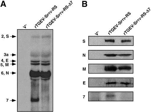
Characterization of mRNAs and proteins of rTGEV-Δ7. (A) Northern blot analysis of viral mRNAs. Total RNA was isolated from BHK-pAPN cells infected with either rTGEVSptv-RS or rTGEV-Sptv-RS-Δ7. V−, noninfected BHK-pAPN cells. The position of viral mRNAs is indicated to the left. (B) Western blot analysis of lysates from BHK-pAPN cells infected with either rTGEV-Sptv-RS or rTGEV-Sptv-RS-Δ7 viruses. Cell extracts were obtained at 16 h p.i., resolved by 5 to 20% gradient SDS–PAGE, transferred to nitrocellulose membranes, and immunoblotted with monoclonal antibodies specific for S, N, M, and E, and with an antiserum specific for a protein 7 peptide (Garwes et al., 1989).