
Fig. 5.
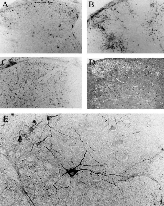
Immunocytochemical staining of the spinal cord of a C57BL/6 mouse (21 DPN) 1 week after HCoV-OC43 infection. (A, B, and C) Evidence of inflammatory reaction on three consecutive sections of the cervical dorsal horn. (A): Antiviral MAb. (B) Mac-2 microglia/macrophages cell marker. (C): GFAP astrocytic cell marker. (D): spongiosis aspect of gray matter revealed by toluidine blue staining. (E): Infection of motor neurons by HCoV-OC43 in the ventral horn of the lumbar spinal cord. Magnification: ×100 before enlargement.