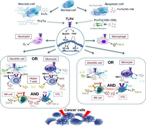
Fig. 2.

Proposed scenario on proTα’s dual role. In normal cells, proTα regulates gene expression and cell proliferation in the nucleus. Under abnormal conditions, cells die via necrosis or apoptosis. During necrosis, cell membrane is ruptured and intact proTα is released out of the cell. During apoptosis, proTα is relocated to the cytoplasm and is cleaved at its C-terminus by activated caspases, and proTα(100–109) is generated. The decapeptide adopts a β-sheet conformation and is excreted. Extracellularly, both proTα and proTα(100–109) activate innate immune cells expressing TLR4 (macrophages, neutrophils, DCs, and monocytes) and signal through the MyD88 and TRIF pathways. Cytotoxic responses are enhanced through antigen presentation with MHC class II molecules and synapsis with helper T cells which secrete TH1-type cytokines, and/or with MHC class I molecules, leading to activation of cytotoxic effectors (NK cells and CTLs). In both cases, effector cells upregulate adhesion molecule expression (eg, CD2) and produce lytic molecules (eg, perforin), mediating cell binding and cell destruction, respectively (eg, cancer cell targets, as shown here).
