
Fig. 2.
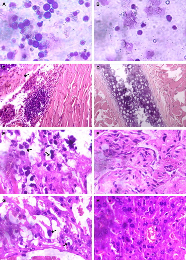
Histopathological analysis of tissues from SCID mice bearing HL-60 cells. Marrow from the thighbone of the mice was obtained, and samples were smeared onto glass microscope slides and stained with Wright's stain. Tissues from the liver and bone were fixed in 4% formaldehyde, embedded in paraffin, sectioned, and stained with hematoxylin and eosin. A: marrow of control, typical leukemia cells could be seen, ×1000; B: marrow of lycorine- (10 mg/kg/day) treated group, ×1000; C: bone of control, the marrow cavity of the sternal bone changed to sawtooth (arrows), ×150; D: bone of lycorine- (10 mg/kg/day) treated group, ×150; E: sides parenchyma of bone in the control group, leukemia cells infiltrate (arrows), ×600; F: sides parenchyma of bone in lycorine- (10 mg/kg/day) treated group, ×600; G: liver of control, leukemia cells infiltrate the liver (arrows) ×600; and H: liver of lycorine- (10 mg/kg/day) treated group, ×600.