
Fig. 2.
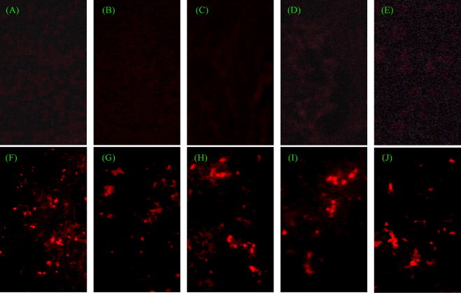
shRNA-expressing plasmids significantly reduced TGEV antigen in different organs. All sample frozen tissue sections were stained with Texas Red (original magnification, 400×). Mesenteric lymph node (A), liver (B), jejunum (C), ileum (D) and kidney (E) were sampled from the mini-pig in group 4. Red spots indicated the TGEV-infected cells. Mesenteric lymph node (F), liver (G), jejunum (H), ileum (I) and kidney (J) were sampled from the mini-pigs in group 2, served as the viral infection controls.