
Fig. 2.
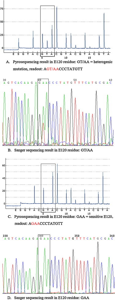
Detection of E120 using the primer S323 and the cyclic dispensation order (GTAC)10. Pyrogram sequence results at residue E120 (highlighted) had the sequence (GT/AA, red color), indicating that glutamic acid (GAA) was partly replaced by valine (GTA) in the specimen isolated from MDCK cells (A) while Sanger sequencing results showed a mix of nucleotides with T/A coding for the amino acid at position 120 (highlighted) in the same specimen (B). Pyrogram sequence results at residue E120 (highlighted) representing the oseltamivir-susceptible codon (GAA, red color) (C) and Sanger sequencing results showed GAA coding for the amino acid at position 120 (D) in its original clinical specimen. The letters listed below the peaks of each panel are the nucleotides dispensed during pyrosequencing on the PyroMark ID system. (For interpretation of the references to color in this figure legend, the reader is referred to the web version of this article.)