
Figure 3.
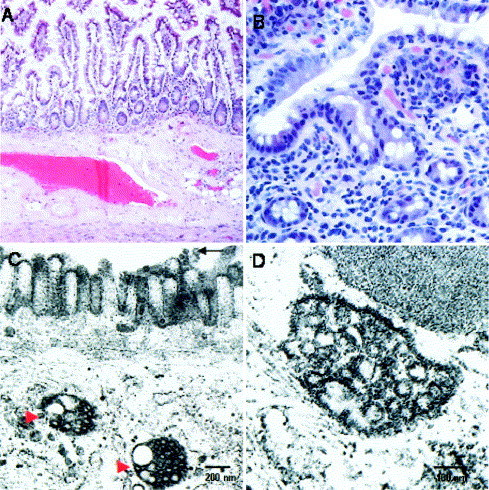
Histologic and ultrastructural appearances of the small intestine in patients with SARS-CoV infection. (A) Section of the small intestine of an autopsy specimen with unremarkable mucosa, submucosa, and muscle layer (H&E; original magnification 40×). (B) Endoscopic ileal biopsy specimen with no inflammatory process (tangentially sectioned, H&E; original magnification 200×). (C) Dilated cytoplasmic vesicles filled with viral particles in the small intestine (indicated by red arrowheads). Scattered viral particles were also detected on the surface microvilli of this surface enterocyte (indicated by black arrow). (D) Higher magnification of a virus-containing vesicle.