
Fig. 5.
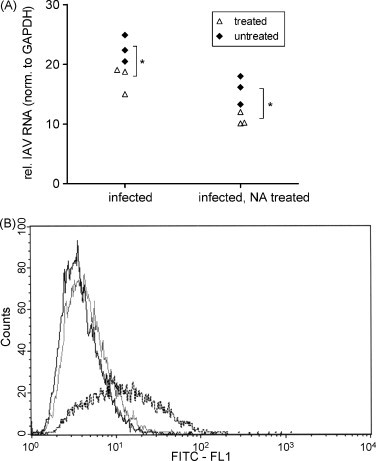
Inhibition of virus uptake. (A) MDCK cells were pre-treated with GL (2.5 mM) before synchronised infection with IAV at MOI 100 and GL (2.5 mM) was present during virus adsorption and uptake. Viral RNA in cell lysates was analysed after virus uptake at 37 °C for 1 h. Surface bound virions were removed after virus uptake at 37 °C for 1 h by NA treatment for 90 min on ice. *p < 0.05. (B) FACS diagram of MDCK cells infected with fluorescence labelled IAV at MOI 10 treated or not with 2.5 mM GL (treatment protocol F) (black line, MDCK cells infected with unlabelled IAV; gray line, MDCK cells GL treated (2.5 mM) infected with fluorescence labelled IAV; dashed line, MDCK cells untreated, infected with fluorescence labelled IAV). (For interpretation of the references to color in this figure caption, the reader is referred to the web version of the article.)